9 months: A guide to prenatal appointments
Posted on
OK, you’ve found out you’re pregnant. If it’s your first baby, it’s safe to say you’ll be venturing into unknown territory.
One thing is certain, however: for the next nine months, you’ll be facing a series of prenatal medical appointments. But what exactly is involved and what does it all mean? With the help of National Capital Diagnostic Imaging (NCDI), we’ve put together this handy guide to break it all down for you.
The Dating Scan
The dating scan is a diagnostic scan performed in the first trimester of pregnancy and provides both the gestational age of your baby and an estimated due date. This scan is best performed at seven to eight weeks gestational age. By this stage, your baby has developed a foetal heart rate and there is less variance in measurement from head to rump.
Preparing for the scan
You will have to fill your bladder by drinking 600mls of water two hours before your appointment. Yes, it’s a bit uncomfortable – but a full bladder will provide a “window” to the uterus. Without water in the bladder, the sonographer is unable to assess the baby and pelvic structures optimally.

What happens on the day of your scan?
Once you have registered you will be asked to take a seat and the sonographer will call your name, introduce themselves and lead you to the ultrasound room. (The sonographer will check with you to make sure you have followed the required preparation.)
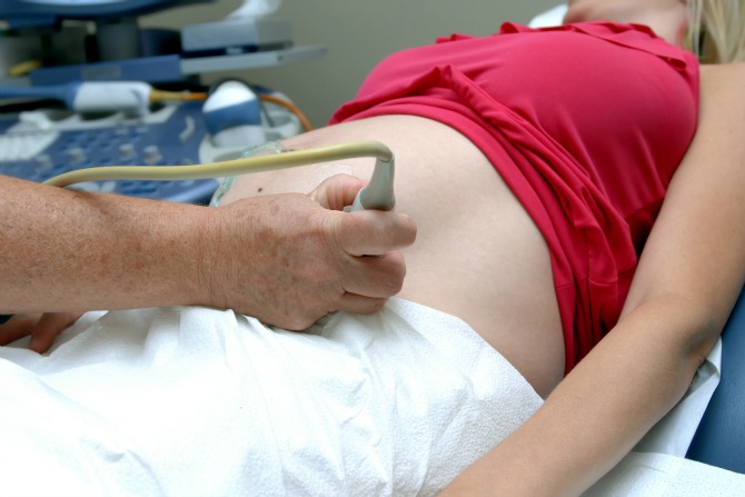
You will then be asked to change into a gown and lie on the ultrasound bed, where gel is placed on your lower abdomen. Chief Sonographer at NCDI Margaret Marchese says she finds the first thing parents want to see is the heart beating. “Seeing this relaxes parents for the rest of the study, making it a more enjoyable experience,” Margaret says.
At the dating scan the sonographer is looking at taking measurements of the baby, the heart rate and the gestational sac. They will also assess your ovaries and uterus, and perform a general survey of the pelvis along with answering any clinical questions raised by the referring doctor. At the seven-eight week mark, your baby will measure approximately 1-2mm and resemble a jellybean.
What if the sonographer is unable to assess the baby optimally through the bladder?
In some instances the baby cannot be assessed optimally through the bladder. In this case an internal examination would be best. This study is known as a transvaginal ultrasound. If this is required, the sonographer will explain the procedure to you and will obtain your consent before proceeding. This examination is performed with an empty bladder and a “modesty sheet” placed over you. The internal examination allows greater visualisation of your baby as the ultrasound beam is much closer, not having to pass through skin, fat, muscle, or bowel gas which all deteriorates the image quality when scanning through the abdomen.
What happens after the scan?
Upon completion of the scan, the sonographer will ask if there are any further questions to ensure your expectations are met.
A doctor (radiologist) will then review the images, ensuring a thorough diagnostic examination has been performed. They will answer any clinical questions raised by your doctor and if necessary provide a diagnosis. The completed report is then forwarded to your referring doctor. NCDI can also provide you with a CD of your pictures along with a printout at this stage. (Please request this from the sonographer.)
Finally, if you have considered having a First Trimester Screening test (Nuchal Translucency test), a blood form is given to you to be performed at the 10 week gestational mark.
The First Trimester Screen (12 week scan)
The First Trimester screen, more commonly known as a Down Syndrome test or Nuchal Translucency test, is to provide a risk assessment of Trisomy 21, 18 and 13 of baby. The measurements required at this stage are the head/crown to rump measurement, the heart rate, and the nuchal translucency.
To perform this scan, the baby’s length from head to rump must be in a particular range, ideally when your baby is 12 weeks or greater as it allows the sonographer to also make an assessment of baby’s anatomy such as the development of the skull which isn’t so easily visualised before 12 weeks.
At this stage of the pregnancy, your baby has developed arms, legs, hands and feet, all which you will see moving. Baby can move all around the gestational sac, rolling onto its tummy or even standing on its head. Sonographers who perform these studies are required to undertake specific additional training.
Preparing for the scan
To increase the detection rate of the test, this test is best performed in conjunction with a blood test (maternal serum screening). If the blood test has been performed prior to the ultrasound, then the complete risk assessment can be provided to your GP for you to discuss within 24hours.
For this scan, the sonographer will also require you to have a full bladder for the examination to better visualise your baby.
What happens on the day of your scan?
You will be asked to answer a questionnaire at the time of the examination asking such questions as height and weight to help calculate a risk assessment as accurately as possible.
To take the Nuchal measurement, the sonographer requires baby to lie in a position which meets all the criteria.
The sonographer will make a full assessment of the uterus and pelvis, taking note of such things as the placenta. A mini anatomy scan is performed of baby, noting such things as hands, feet, nasal bone, bladder etc.
20 Week Morphology Scan
At this stage of the pregnancy, your baby has developed all the relevant anatomy for the sonographer to see, it simply needs to grow in size and weight.
Baby may not be able to fit across the screen as it did at the 12 week scan, but you will see movement such as swallowing, yawning, touching toes and even extending feet over its head.
The 20 week morphology scan is performed to assess the anatomy of baby such as face and head, arms and legs, spine, kidneys, bladder and the heart. The reason the scan is best performed at the 20 week mark is to better visualise and assess the heart. The heart at this stage is the size of a thumb nail and therefore the bigger it is, the easier it is to see.
Measurements will be performed on baby to see if it is growing accordingly with your estimated date of delivery. A heart rate is also recorded.
The sonographer will also again access the uterus, fluid around baby, placental location, cervix and surrounding maternal structures.
Preparing for the scan
Make sure you are well hydrated so that the sonographer can more easily see the cervix.
What happens on the day of your scan?
The sonographer will not ask if you would like to know the gender of the baby as the focus is on performing a diagnostic scan of baby’s anatomy. If you would like to know the gender of your baby, please ask the sonographer to check at the beginning of the exam, that way they have plenty of time to be sure of what the gender is.
Other information you should tell the sonographer is if you have any family history or previous pregnancies with any structural anomalies, any previous surgeries to your cervix and if any scans have been performed elsewhere.
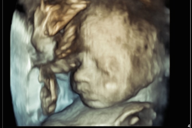
The scan time can vary depending on how baby is positioned in the uterus and the difficulty of the scan. The NCDI can allow one hour appointments so that the sonographer has enough time to perform a thorough examination without any time constrains. The focus is on providing a positive experience, allowing time to see your baby move around on the screen. NCDI do have the capability to perform a 3D image at selected clinics. This is however dependant on the amount of fluid overlying baby’s face, placental location and baby’s position and may not be possible with all patients.
With regards to bringing additional family members into the room, NCDI prefer this appointment to be limited to one other person to avoid too much distraction. The radiologist will review the scans and provide a detailed report commenting on such things as if the placenta is clear of the internal os (cervix), the fluid levels around baby, the growth measurements of baby along with baby’s approximate weight and if any anomaly was seen.
A CD of your pictures along with a printout can also be provided.
Third Trimester Scan
This scan is performed in the third trimester (greater than 27 weeks) if further investigation is required or requested by your specialist.
Preparing for the scan
The preparation again is to be well hydrated and it is very important to bring along any previous films or reports to the scan.
What happens on the day of your scan?
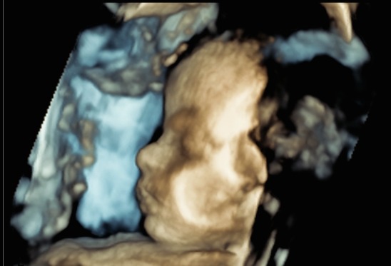
Your baby has grown considerably at this stage, and it becomes more difficult to see its face clearly. The sonographer may be able to point out the profile if there is fluid overlying baby’s face, or it might be easier to point out the lips and nose.
The sonographer will tailor the examination to your doctor’s request, with a focus on answering the clinical questions raised. Routine measurements are performed of baby again and plotted against a growth chart to see if baby is growing accordingly to dates and in which percentile baby is growing (between 5-95 per cent).
Assessment is also made of the fluid around baby, the placenta, the cervix, and doppler blood flow measurements are obtained of the umbilical cord and if necessary the Middle Cerebral Artery in baby’s brain. A heart rate is obtained and the sonographer will observe the movement of baby over time, seeing if baby is practising it’s breathing and looking at other gross and fine movements.
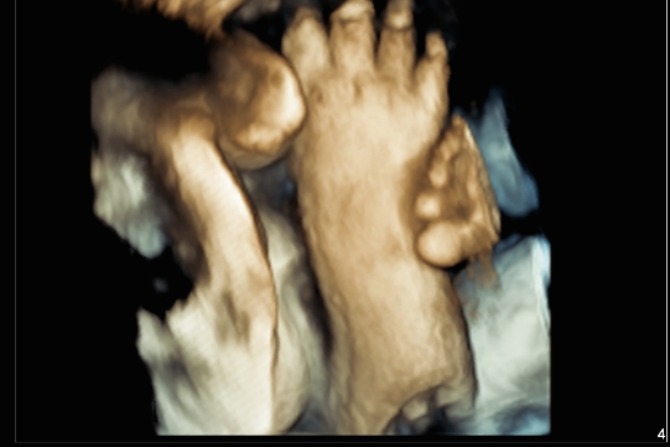
This also gives an opportunity for the sonographer to show the mum how baby moves, the opening and closing of baby’s hands or taking a big yawn. You may even see a foot pressed up against mum’s belly.
A more basic anatomy scan is also performed looking at kidneys, bladder diaphragm, etc.
The sonographer may ask you how you have been going throughout the pregnancy to obtain any relevant clinical information such as blood pressure.
The radiologist will again provide a report commenting on the placenta location, growth of baby, in which percentile baby is in along, the Doppler blood flow measurements and fluid levels making sure they are within normal range. The report will also state the position baby is lying in as it can determine how you deliver.
About National Capital Diagnostic Imaging (NCDI)
National Capital Diagnostic Imaging in Tuggeranong, Woden and Deakin, offers a comprehensive range of medical imaging services and scans including x-ray, CT, MRI, ultrasound, obstetric ultrasound, mammography, nuclear medicine and interventional procedures.
NCDI has provided quality medical imaging services to the ACT community for over 30 years and take great pride in providing patients with a caring, professional and clinically superior service.
NCDI is sensitive to the intimate nature of women’s imaging, and doctors and staff are committed to providing the most accurate diagnosis possible in a caring and sensitive environment focused on something increasing forgotten these days – your comfort.
Where to go
Deakin
Canberra Specialist Centre Suites A2 & B2, 161 Strickland Crescent Deakin, ACT 2600
6124 1900
Tuggeranong
Suite 10a, Homeworld Centre Anketell Street Tuggeranong, ACT 2900
6293 2922
Woden
Woden Specialist Medical Centre
90 Corinna Street Woden, ACT 2606
Ground Floor: 6214 2222
Level 1: 6122 7878
This is a sponsored post. For more information on our sponsored post policy, click here.
Images used throughout the article provided by NCDI. Feature image: Shutterstock


Leave a Reply
You must be logged in to post a comment.